November 14, 2015 Admin Home page » Lat + upper back
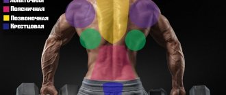
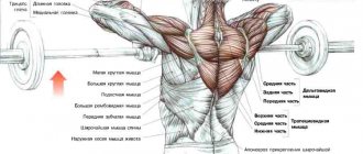
The anatomy of the back muscles is described, each muscle is shown in detail with a photo, find out what our back is made of, to strengthen it and create sculpted muscular muscles.
Anatomy of the back muscles - a huge, wide back always distinguishes ordinary people from bodybuilders, the famous “triangular frame” is achieved only through the development of this muscle group, and for a girl, a beautiful, slender posture, the standard and main advantage of her gait.
Powerful and strong back muscles are used in almost all sports, as they take part in all pushing, pulling and twisting movements, also with strong back muscles, our spine supports a living shield of “flesh and meat”. Especially strong lower back muscles will allow you to get rid of the feeling of pain when bending over or lifting moderately heavy things.
Superficial muscles
The back muscles attached to the bones of the shoulder girdle are called superficial. They are arranged in two layers .
Layer one
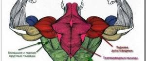
M. trapezius, trapezius muscle
It resembles a flat figure, the upper side of which forms the lateral cervical triangle, the lower lateral side crosses the scapula laterally. Insertion points: acromial end of the clavicle, acromion, spine of the scapula.
It starts from the occipital protrusion, from the edge of the nuchal occipital bone, ligaments - nuchal, supraspinous, CVII, thoracic vertebrae. Contraction of all parts of the muscle pulls the scapula towards the spine . Upper beams m. trapezius raise the scapula, the lower ones lower it. Together, rotate the scapula in a vertical plane.
Latissimus muscle, m. latissimus dorsi
The upper side of the muscle triangle is hidden under the lower part of the m. trapezius. The lower one forms the lateral side of the lumbar triangle. The powerful muscle responsible for adducting, turning inward, lowering the arm, pulling the torso towards the arms (when swimming, climbing, pulling up) has a solid origin.
From all lumbar, spinous processes of the lower 6 thoracic vertebrae. From the median sacral and iliac crests. Additionally from above to m. latissimus dorsi muscle bundles are attached, starting from the lower 4 ribs and the angle of the scapula. The crest of the lesser tubercle of the humerus is the site of attachment.
Video: “Classification of back muscles”
Second layer
Mm. rhomboidei minor et major - small and large rhomboid muscles
They can merge into one. M. rhomboidei minor starts from the lower part of the nuchal ligament, spinous processes of CVII, TI, and supraspinous ligament. Large - from the spinous processes of TII - V. The place of muscle attachment is the lateral edge of the scapula.
Only m. rhomboidei minor is attached above the level of the spine, and major - from the level of the spine to the lower angle of the scapula. M. rhomboidei are located deeper than m. trapezius. They bring the scapula towards the spine, moving upward .
M. levator scapulae, levator scapulae muscle
Performs the function according to its name . At the same time, it brings the scapula closer to the spine and tilts the neck (with the scapula fixed). Start m. levator scapulae - posterior tubercles of the transverse processes of the 4th or 3rd upper cervical vertebrae. Attached to the side of the shoulder blade.
The serratus posterior muscles stand apart in the classification.:
- M. serratus posterior superior - the superior posterior serratus muscle is responsible for raising the ribs. It is located in front of mm. rhomboidei. Origin - the lower part of the nuchal ligament, spinous processes of the VI-VII cervical, I-II thoracic vertebrae. Attachment - posterior angular surface of ribs II-V, with separate teeth.
- M. serratus posterior inferior - the lower posterior serratus muscle lowers the ribs . Lies in front of m. latissimus dorsi. Starts from the spinous processes ТXI – XII, LI-II. It is attached to the 4 lower ribs with separate teeth.
Video: “Superficial back muscles”
Deep muscles
The deep back muscles are represented in three layers.
Surface
M. splenus capitus - splenius capitis muscle
Extends the neck and head, contracting on both sides . Turns his head, contracting on one side. Origin: nuchal ligament below CIV, CVII, upper (3rd or 4th) thoracic vertebrae. The muscle bundles pass upward and are laterally attached to the mastoid process of the temporal bone and the rough area under the lateral segment of the superior nuchal line of the occipital bone.
M. sprlenius cervicis - splenius muscle of the neck
Extends the neck while simultaneously contracting . Unilateral contraction of the muscle turns the neck. Starts from the spinous processes TIII-IV. It is attached to the posterior tubercles of the transverse processes of the 3 upper cervical vertebrae. Located in front of m. trapezius.
M. erector spinae, erector spinae muscle - strengthened
Responsible for holding the body in an upright position . Stretches from the sacrum to the base of the skull. Triples at lumbar level – m. iliocostalis (iliocostal), m. longissimus (longest), m. spinalis (spinous). Attached to the ribs, vertebrae, and base of the skull.
Average
M. Transversospinalis - transverse spinalis muscle
Responsible for straightening the body, bending, and turning the spine . The muscle is divided into parts that spread across different groups of vertebrae - m. semispinalis (semispinalis), mm. multifidi (multipartite), mm. rotatores (rotators). These muscle groups rotate the spine, move the head, and control breathing movements.
Deep
Mm. interspinales cervicis, thoracis et lumborum - interspinous muscles of the neck, chest and lower back
Participate in the extension of the departments corresponding to the name . The spinous processes unite each other. Down from the second cervical vertebra. These muscles are poorly developed in the thoracic spine or may be absent.
Mm. intertransversarii lumborum, thoracis et cervicis - intertransverse muscles of the lower back, chest, neck
Responsible for the inclination of the homonymous sections of the spinal column . These are short muscle bundles between the transverse processes of adjacent vertebrae.
Mm. Suboccipitales - suboccipital muscles
They are located deep under the superficial muscles. They enclose the vertebral artery, the posterior branches of the spinal nerve CI, the arch of the atlas and the atlanto-occipital membrane in a triangular space. Responsible for head movements - throwing back, tilting, rotating .
Video: “Deep back muscles”
Back training - beginner level
This training program is designed for those who are just starting their journey to a wide and strong back.
This routine consists of back exercises in the gym targeting all the major parts of the upper and lower region, including the lats, trapezius, rhomboids, teres muscles, and rear deltoids.
The volume of these exercises is specially selected so that you can pump up all the key muscle groups, but the next day you won’t collapse from exhaustion. After all, the key to high-quality muscle growth is adequate load during training and good recovery after its completion.
The first two approaches should be done with light weight until the so-called “pre-exhaustion”. That is, if you try to perform a movement, your muscles simply will not be able to complete it.
Perform the last approach until “failure” - this means until the moment when you cannot complete the next repetition at all, no matter how hard you try.
And don’t be shy about asking for backup for maximum safety during training.
Back thickness exercises for beginners:
- Deadlift: 3 x 4-8
- Wide grip seated vertical block row: 3 x 8-15
- Straight-grip lat pull-down: 3 x 8-15
- Upper block row with straight arms: 3 x 8-15
- One-arm dumbbell row: 3 x 8-15
What diseases of the back muscles are there?
Did you know that...
Next fact
There are many names for muscle diseases. Almost always they correspond to the mechanism of the pathological process. In addition to the name “myalgia,” which refers to muscle pain due to overexertion.
Inflammation of the muscles after hypothermia, infection, manifested by pain - myositis . An adhesive fibrotic process in the muscles, a consequence of advanced myositis - fibromyositis . Adhesions between muscle tissues due to thickened lymph released and accumulated around the inflammatory focus - neurofibromyositis .
The blood vessels that innervate and supply the muscle become fused with it during inflammation. Therefore, any influence on the muscle - temperature, static tension causes pain in it.
Muscle pain in the lumbar and sacral region may be a reaction to hysteria. The symptom of herpes zoster is unilateral lower back pain. Any functional changes in the spine are reflected in the condition of the muscles, which try to compensate for the incorrect position of the spinal column by straining.
A constant pathological effect on the muscle forms trigger (sensitive) points that trigger an inadequate pain response at the level of the central nervous system.
Types of myositis and fibromyositis:
Lumbago (myositis, fibrositis) is a muscle disease at the level of the TXII, LV vertebrae . The pathology is caused by a convulsive spasm of the deeply embedded intervertebral muscles. Trigger areas: medial edge of the erector erector muscles, iliac crest, sacroiliac joint.
A characteristic symptom, in addition to pain, is scoliosis in the affected area, low-grade fever. Lumbago must be differentiated from a tumor of the spinal cord and spine, myelitis. If the patient has muscle pain, fever, eosinophilia, diarrhea, swelling of the skin, then trichinosis cannot be ruled out.
Myositis of the lumbar muscles has a long-term cure with passive treatment. Its chronic form does not manifest itself with such severe pain as with lumbago. The pain is aching and irritating to the patient. Adjoining fibrositis maintains the pain syndrome.
- Dermatomyositis , accompanied by skin changes and pain, is caused by autoimmune changes. The disease is sometimes mistaken for scleroderma, lupus erythematosus, or polyneuritis. Pathology may be a symptom of malignant tumors.
– is always secondary. This is a consequence of an infection during injection, a muscle abscess.
Purulent myositis- Myositis ossificans - calcification of areas of injured muscles. Muscles that have lost their elasticity become very painful. Body temperature rises.
- Toxic myositis is observed in chronic alcoholics or those exposed to intoxication with pharmacological agents. Accompanied by paresis and muscle swelling.
- Neuromyositis – changes in muscle nerve fibers, distal axons of nerves. Severe pain that increases with palpation. Balle's painful points. Mild symptoms of tension.
- Polyfibromyositis is accompanied by pain during movements, thickening of muscles in the attachment zone, and contractures. The muscles do not relax during sleep or during general anesthesia.
- Acute alimentary myositis or Cox-Sartlan disease. The disease is associated with the consumption of certain types of fish. Its epidemic outbreaks occur in fishing villages. Myorenal syndrome is characteristic. Toxins act simultaneously on the muscular system and kidneys. Symptoms are sharp pain in the lower back, legs, arms, chest. The patient has difficulty breathing, hyperhidrosis, vomiting, and blood in the urine.
Pathological conditions causing pain in the back muscles due to traumatic injury, overexertion, hypothermia:
- Ligament sprain . Causes: falls, sports injuries, heavy lifting. The longitudinal and interspinous ligaments of the spine are damaged. Most often the area TVII – TVIII.
- Torticollis . Torsion dystonia with involuntary contraction of the neck muscles is manifested by rotational deformity. It can be congenital or acquired.
- Scapular-costal syndrome – damage to m. levator scapulae. Pain in the neck, shoulder blade, shoulder joint. The trigger zone is where the muscle attaches. When moving the blade, a crunching sound is heard. The disease affects the levator scapulae muscle, adjacent muscles, fascia, and ligaments.
- Myofasciculitis of the trapezius muscle . In case of injury or hypothermia, it has an acute onset. With the development of burning, boring pain at the slightest movement in the cervical and thoracic region. On palpation at the site of muscle attachment there is an acute pain response. The patient has difficulty choosing a position in bed. Prolonged exposure to low temperatures can cause gradual development of pathology. It is accompanied by a feeling of heaviness along the muscle, a dull aching pain that intensifies with static and dynamic loads and radiates to the intercostal spaces. Small, painful lumps may migrate under the skin during massage. Painful manifestations of myofasciculitis are often confused with heart pain.
- Myofasciculitis of the rhomboid muscles . Pain in the area of the dorsum of the forearm increases gradually. Intensifies when turning the head and arms. By palpation, pain is determined at the medial edge of the scapula and spinous processes.
- Myofasciculitis of the serratus muscle . Manifestations: heaviness, cerebral (dull, aching) pain at the CV-CVII level. Intensifies at night after exercise.
- Myofasciculitis m. latissimus dorsi . The symptoms are similar to those listed above.
Video: “10 facts about the back muscles”
Erector spinae muscles: how to train and relax
A feature of these muscles is their slow recovery. Therefore, it is often not recommended to strain them. It is better to train with strength exercises no more than 2 times a week. The rest of the time, your classes should include exercises to relax and stretch these muscles. This will help relieve their spasm:
- The simplest exercise to relax the back muscles is hanging on a horizontal bar. It is recommended to stay in this position for several minutes 2-3 times a day.
- Sit on a chair, spread your legs wide, lower your arms. Exhaling slowly, alternately bend the spine in the cervical, thoracic and lumbar regions, drawing in the stomach. As you inhale, straighten up, straightening your back in the reverse order.
- Lie on your back, clasp your hands around the knees of your bent legs. As you inhale, press your legs on your arms, as if trying to straighten them, exhale - bring your knees closer to your head.
