Vintage drawing of human muscles
Muscles
or
muscles
(from Latin musculus - muscle) - a part of the musculoskeletal system in conjunction with the bones of the body, capable of contraction. Designed to perform various actions: body movement, maintaining posture, contraction of the vocal cords, breathing. Muscles consist of elastic, elastic muscle tissue, which, in turn, is represented by cells called myocytes (muscle cells). Muscles are able to contract under the influence of nerve impulses. Muscles are characterized by fatigue, which manifests itself during intense work or stress.
Muscles allow you to change the position of body parts in space. A person performs any movement - from such simple movements as blinking or smiling, to subtle and energetic ones, such as we see in jewelers or athletes - thanks to the ability of muscle tissue to contract. Not only the mobility of the body, but also the functioning of all physiological processes depends on the proper functioning of the muscles, consisting of three main groups. The work of all muscle tissue is controlled by the nervous system, which ensures their connection with the brain and spinal cord and regulates the conversion of chemical energy into mechanical energy.
There are 640 muscles in the human body (depending on the method of calculating differentiated muscle groups, their total number is determined from 639 to 850) [ source not specified 2010 days
]. The smallest ones are attached to the smallest bones located in the ear. The largest muscles are the gluteus maximus muscles, they move the legs. The strongest muscles are the calf muscles and the masseter muscles. The longest muscle in humans, the sartorius, starts from the anterior superior bone of the iliac wing (anterior superior parts of the pelvic bone), spirals across the front of the thigh and is attached by a tendon to the tuberosity of the tibia (upper parts of the tibia).
The shape of the muscles is very diverse. The most common are the fusiform muscles, characteristic of the limbs, and the vastus muscles, which form the walls of the torso. If muscles have a common tendon and two or more heads, then they are called two-, three-, or four-headed.
Muscles and skeleton determine the shape of the human body. An active lifestyle, a balanced diet and exercise help develop muscles and reduce the volume of adipose tissue. Muscle mass among leading weightlifters accounts for 55-57% of body weight[1].
Muscle types
Types of muscle tissue (by structure): skeletal, smooth and cardiac
Additional information: Skeletal muscle tissue, Smooth muscle, and Cardiomyocyte
Depending on the structural features, human muscles are divided into 3 types or groups:
- skeletal,
- smooth,
- cardiac.
The first muscle group is skeletal
, or striated muscles. Each of us has more than 600 skeletal muscles. Muscles of this type are capable of contracting voluntarily, at the request of a person, and together with the skeleton form the musculoskeletal system. The total mass of these muscles is about 40% of body weight, and in people who actively develop their muscles, it may be even more. With the help of special exercises, the size of muscle cells can be increased until they grow in mass and volume and become defined. As a muscle contracts, it shortens, thickens, and moves relative to neighboring muscles. The shortening of the muscle is accompanied by the convergence of its ends and the bones to which it is attached. Each movement involves muscles both performing it and opposing it (agonists and antagonists, respectively), which gives the movement precision and smoothness.
The second type of muscle, which is part of the cells of internal organs, blood vessels and skin, is smooth muscle tissue.
, consisting of characteristic muscle cells (myocytes). Short spindle-shaped smooth muscle cells form plates. They contract slowly and rhythmically, obeying signals from the autonomic nervous system. Their slow and prolonged contractions occur involuntarily, that is, regardless of a person’s desire.
Smooth muscles, or muscles of involuntary movement, are found mainly in the walls of hollow internal organs, such as the esophagus or bladder. They play an important role in processes independent of our consciousness, such as the movement of food through the digestive tract.
A separate (third) group of muscles is the cardiac striated (striated) muscle tissue (myocardium). It consists of cardiomyocytes. Contractions of the heart muscle are not controlled by human consciousness; it is innervated by the autonomic nervous system.
Muscle functionality
We don’t really think about how the muscles work during our movements; for us, all these actions happen automatically. The human body is a well-established mechanism, each element of which has its own functionality.
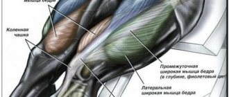
Quadriceps
Works with knee extension. Its constituent rectus muscle bends the body at the waist to an angle of 90 degrees, while it works separately from the other muscles.
Back of thigh
Basically, this muscle group works in the same way, namely: it extends at the hip joint and bends at the knees. Extends the body in the pelvic region, if the limb is fixed, and acts together with the gluteus maximus muscle. When the knee is bent, it allows for inward and outward rotation of the lower leg.
Buttocks
They function by bringing the hips together, and also perform rotational movements in and out. If the limbs are fixed, then the body is extended and the pelvis is tilted to the sides.
Adductors
They are also called adductors or “moral muscles.” In addition to their key role in hip adduction, they are involved in pelvic flexion and extension and axial rotation of the limb. Another important feature is that they help stabilize the body. The training of these muscles helps to quickly maneuver, which is especially important during active sports.
Posterior surface of the leg
In addition to bending the knee and foot, the muscles here twist the lower leg, supinate the foot, bend the toes, and press them toward the ground while standing.
Structure
| The information in this section is out of date. You can help the project by updating it and then removing this template. |
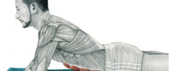
The structure of skeletal muscle
The structural element of muscles is muscle fiber, each of which individually is not only a cellular, but also a physiological unit capable of contraction. The muscle fiber is a multinucleated cell, its diameter ranges from 10 to 100 microns. This cell is enclosed in a membrane, the sarcolemma, which is filled with sarcoplasm. Myofibrils are located in the sarcoplasm. Myofibril is a thread-like formation consisting of sarcomeres. The thickness of myofibrils is generally less than 1 µm. Depending on the number of myofibrils, white and red muscle fibers are distinguished. White fibers have more myofibrils and less sarcoplasm, so they can contract more quickly. Red fibers contain a large amount of myoglobin, which is why they got their name. In addition to myofibrils, the sarcoplasm of muscle fibers also contains mitochondria, ribosomes, the Golgi complex, lipid inclusions and other organelles. The sarcoplasmic reticulum ensures the transmission of excitation impulses within the fiber. Sarcomeres are composed of thick myosin filaments and thin actin filaments[2].
Actin
is a contractile protein consisting of 375 amino acid residues with a molecular weight of 42300, which makes up about 15% of muscle protein. Under a light microscope, thinner actin molecules appear as a light stripe (the so-called “Ι-discs”). In solutions with a low ion content, actin is contained in the form of single molecules with a spherical structure, but under physiological conditions, in the presence of ATP and magnesium ions, actin becomes a polymer and forms long fibers (fibrillar actin), which consist of two spirally twisted chains of actin molecules. By combining with other proteins, actin fibers acquire the ability to contract using the energy contained in ATP.
Myosin
- main muscle protein; its content in muscles reaches 65%. The molecules consist of two polypeptide chains, each containing more than 2000 amino acids. The protein molecule is very large (these are the longest polypeptide chains existing in nature), and its molecular weight reaches 470,000. Each of the polypeptide chains ends in a so-called head, which includes two small chains consisting of 150-190 amino acids. These proteins exhibit the enzymatic ATPase activity required for actomyosin contraction. Under a microscope, myosin molecules in muscles appear as a dark stripe (the so-called “A-discs”).
Actomyosin
- a protein complex consisting of actin and myosin, characterized by the enzymatic activity of ATPase. This means that thanks to the energy released during the hydrolysis of ATP, actomyosin can contract. Under physiological conditions, actomyosin creates fibers that are in a certain order. The fibrillar parts of myosin molecules, collected in a bundle, form a so-called thick filament, from which the myosin heads protrude perpendicularly. Actin molecules are connected into long chains; two such chains, spirally twisted around each other, make up a thin thread. Thin and thick threads are arranged in parallel in such a way that each thin thread is surrounded by three thick ones, and each thick thread by six thin ones; myosin heads cling to thin filaments.
In general, muscle tissue consists of water, proteins and a small amount of other substances: glycogen, lipids, extractive nitrogen-containing substances, salts of organic and inorganic acids, etc. The amount of water is 72-80% of the total mass [2].
Chemical composition of mammalian striated muscles (average values)[2]
| Component | Percentage of wet weight |
| Water | 72—80 |
| Dense substances | 20—28 |
| including | |
| squirrels | 16,5—20,9 |
| glycogen | 0,3—3,0 |
| phosphoglycerides | 0,4—1,0 |
| cholesterol | 0,06—0,2 |
| creatine + creatine phosphate | 0,2—0,55 |
| creatinine | 0,003—0,005 |
| ATP | 0,25—0,40 |
| carnosine | 0,2—0,3 |
| carnitine | 0,02—0,05 |
| anserine | 0,09—0,15 |
| free amino acids | 0,1—0,7 |
| lactic acid | 0,01—0,02 |
| ash | 1,0—1,5 |
Muscle proteins are usually divided depending on their solubility in water or saline media. There are three main groups of proteins: sarcoplasmic (35% of the total protein), myofibrillar (45%) and stromal proteins (20%). The composition of sarcoplasmic proteins includes several protein substances that have the properties of globulins, a number of proteins, myoglobin, enzyme proteins, and parvalbumin. Parvalbumin sequesters Ca2+ levels, which accelerates muscle relaxation. Enzyme proteins are located in mitochondria and regulate the processes of tissue respiration, nitrogen and lipid metabolism, etc. Sarcoplasmic proteins dissolve in salt environments with low ionic strength.
Myosin, actin and actomyosin belong to the group of myofibrillar proteins responsible for muscle contraction. This also includes regulatory proteins: tropomyosin, troponin, α- and β-actinin. The complex of tropomyosin and troponin is responsible for muscle sensitivity to Ca2+ ions. Myofibrillar proteins dissolve in saline media with high ionic strength. The content of myofibrillar proteins depends on the type of muscle, while the proteins also differ in their physicochemical properties. The greatest number of them is observed in skeletal muscles, there are much fewer of them in the myocardium, and least of all in smooth muscles. For example, in the muscle tissue of the stomach there are 2 times less proteins of this group than in striated muscles.
Stromal proteins include collagen and elastin. In contrast to myofibrillar proteins, the content of stromal proteins is highest in smooth muscle and cardiac muscle.
As the body develops, a significant change in the chemical composition of muscles occurs. The total protein content in the muscle tissue of embryos is less than that of adults, and, accordingly, there is more water. The composition of the protein mass itself also differs: as development progresses, the amount of stromal proteins decreases and the content of myosin and actomyosin increases. There is also a decrease in the presence of nucleoproteins, RNA and DNA, and the proportion of high-energy compounds (ATP and creatine phosphate) increases. The appearance of certain elements in muscle tissue is associated with specific stages of development. During the formation of the reflex arc and the development of the motor reflex, imidazole-containing dipeptides (anserine and carnosine) appear in the muscles, and Ca2+ sensitivity of actomyosin is formed [2].
The structure of muscle fiber and the mechanism of muscle function
A muscle fiber is a single cell with thin (actin) and thick (myosin) filaments surrounded by mitochondria. The threads are able to interact in small areas of the fibers, this space is called a sarcomere and in total makes up 30% of the length of the muscle fiber, so the muscle can only shorten by 30% of its length. Outside each fiber there is a feeding capillary and a nerve cell process (motoneuron axon); at the point of “connection” to the nerve cell there is a tank containing calcium ions.
The mechanism of muscle contraction (sliding filament theory 1954): at rest, the interaction zone is filled with “brake fluid” - magnesium ions (Mg2+), which allows you not to waste energy at rest. When an exciting impulse passes, calcium ions leave the tank into the interaction zone and remove the “brakes” from actin filaments and activate the centers of myosin molecules, after which contraction occurs. After stimulation ends, calcium returns to the tanks and relaxation occurs.
During muscle work, glucose (glycogen) and fatty acids act as an energy source with sufficient oxygen concentration. Muscles are capable of storing adenosine triphosphate (a source of energy), but these reserves in the muscle are only enough for eight single contractions. To resynthesize ATP, the body uses reserves of creatine phosphate - an energy storage and transmitter from mitochondria to acto-myosin complexes.
Human musculoskeletal system. The growth and development of muscles and bones are closely related - bones are a fulcrum and storehouse of calcium for muscles, and muscles, in turn, regulate the nutrition and growth of bones up to 25 years in length. The muscle is attached by a tendon to the periosteum and, when contracted, stretches it, creating a “subperiosteal space”, the metabolic processes in which are much more intense. This allows the cells to build bone beams more quickly and efficiently, and as a result the bone grows in thickness. This is the main mechanism for strengthening bones, explaining that it is impossible to achieve results only by increasing the concentration of calcium in the blood without accompanying muscle work.
Classification
The muscle tissue of living organisms is represented by numerous muscles of various shapes, structures, development processes, and performing various functions. There are:
By function
- flexors (lat. flexores)
- extensors (lat. extensores)
- abductors (lat. abductores)
- adductors (lat. adductores)
- rotators (lat. rotatores) inside (lat. pronatores) and outside (lat. supinatores)
- sphincters (lat. sphincteres) and dilators
- synergists and antagonists
- lifting (lat. levatores)
- lowering (lat. depressores)
- straightening (lat. erectores)
By grain direction
- rectus muscle - with straight parallel fibers;
- transverse muscle - with transverse fibers;
- orbicularis muscle - with circular fibers;
- oblique muscle - with oblique fibers: unipennate - oblique fibers are attached to the tendon on one side;
- bipinnate - oblique fibers are attached to the tendon on both sides;
- multipinnate - oblique fibers are attached to the tendon from several sides;
- semitendinosus;
- semimembranous.
In relation to the joints
The number of joints through which the muscle is thrown is taken into account:
- single-joint
- two-joint
- multi-joint
By shape
- simple fusiform
- straight long (on limbs)
- short
- wide
- multi-headed two-headed
- square
Muscle contractions
During contraction, actin filaments penetrate deep into the spaces between myosin filaments, and the length of both structures does not change, but only the total length of the actomyosin complex is reduced - this method of muscle contraction is called sliding. The sliding of actin filaments along myosin filaments requires energy ; the energy required for muscle contraction is released as a result of the interaction of actomyosin with ATP with the cleavage of the latter into ADP and H3PO4.
In addition to ATP, water plays an important role in muscle contraction, as well as calcium and magnesium ions.
Skeletal muscle consists of a large number of muscle fibers - the more there are, the stronger the muscle.
There are five types of muscle contractions:
- Concentric contraction - causes shortening of the muscle and movement of its attachment to the bone, while the movement of the limb provided by the contraction of this muscle is directed against overcome resistance, such as gravity.
- Eccentric contraction - occurs when a muscle lengthens while adjusting the speed of movement caused by another force or in a situation where the maximum effort of the muscle is not enough to overcome the opposing force. As a result, movement occurs in the direction of the external force.
- Isometric contraction is an effort that counteracts an external force, in which the length of the muscle does not change and movement in the joint does not occur.
- Isokinetic contraction is the contraction of a muscle at the same speed.
- Ballistic movement is a rapid movement involving: a. concentric movement of agonist muscles at the beginning of the movement; b. inertial movement during minimal activity; V. eccentric contraction to slow movement.
In the body, such contractions are more important for performing any movements.
Smooth muscles (smooth muscle tissue) make up internal organs, in particular the walls of the esophagus, blood vessels, respiratory tract and genitals. Smooth muscles are distinguished by so-called automatism, that is, the ability to enter a state of excitation in the absence of external stimuli. And if the contraction of skeletal muscles lasts about 0.1, then slower contractions of smooth muscles last from 3 to 180 s. In the esophagus, genitals and urinary canal, excitation is transmitted from one muscle cell to the next. As for the contraction of smooth muscles located in the walls of blood vessels and in the iris of the eye, it is not transferred from cell to cell; The sympathetic and parasympathetic nerves of the autonomic nervous system approach the smooth muscles.
Speaking about the heart muscle (myocardium), it should be noted that during normal operation it spends about 0.2-0.4 s to contract, and with increasing load the speed of contraction increases. A unique feature of the heart muscle is its ability to contract rhythmically even when the heart is removed from the body.
During muscle contraction, when actin protein filaments slide along myosin filaments, myosin is temporarily attached to actin with the help of cross bridges, which are the so-called “heads” of myosin molecules. There are 5 stages of the biochemical cycle of muscle contraction[2]:
- the stoichiometric process of ATP hydrolysis by the myosin “head” to adenosine diphosphate (ADP) and orthophosphoric acid (H3PO4); this process does not ensure the release of hydrolysis products;
- binding of the freely rotating myosin “head” containing ADP and H3PO4 to F-actin;
- release of ADP and H3PO4 from the actin-myosin complex;
- binding of the myosin-F-actin complex to a new ATP molecule;
- relaxation stage - separation of the myosin (ATP) “head” from F-actin.
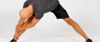
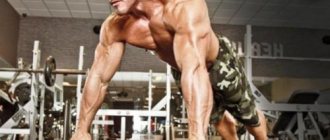
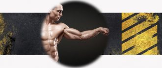
Basic exercises for all leg muscle groups with names and photos
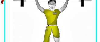
Front Squats
For some reason, recently this species has been unreasonably forgotten. And very much in vain.
First of all, it should be said that it is performed with a barbell on the chest. It is extremely simple to perform and at the same time removes some of the load from the back.
Lunges
They definitely need to be included in the training program. They work the muscles well and are also the least traumatic.
In the first stages, practice in front of a mirror. The feet should be clearly under the hips to create a straight line. As you inhale, take a step forward. The weight should be evenly distributed. Three right angles should form: between the quadriceps and the torso, in the right and left knee. Then - to the starting position.
Simple enough isn't it?
And now, common mistakes:
- Unfixed limb.
- Violation of the correct angle.
- Falling to the surface.
- Rounding the back.
The exercise is very versatile; it can be performed with weights, to the side, or with twisting.
Romanian deadlift
A very effective exercise. Which muscle groups in the legs are involved in the work the most. Gluteal and calf muscles, and also loads the lower back. Where there is benefit, there is also injury. It is extremely important to follow recommendations and safety precautions.
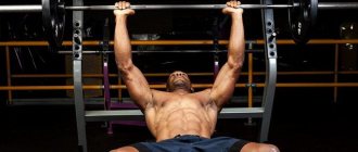
Standing calf raise
Develops mainly calves. You can do it in many variations: at home, in the gym, with dumbbells or a barbell. People with knee joint problems should use it with caution, as it puts a lot of stress on them. Don't forget about preheating and preparation.
Evolution of muscles
An accurate representation of the process of muscle formation over the course of evolution is not yet available. It is known that the first creatures in which the appearance of muscle cells is noted are flatworms and roundworms. Contractile fibers are also present in unicellular organisms, protozoa, and are found in sponges and coelenterates. Contraction of the processes of epithelial cells, vibrations of flagella and cilia allow them to move, but they do not have specialized muscle cells. The musculature of many worms is a so-called musculocutaneous sac, which is formed by muscle fibers separate from the epithelium and associated with the skin. These muscles are close to the smooth muscles of vertebrates and usually consist of external circular fibers, which allow the worms to reduce their diameter, and internal longitudinal fibers, which allow them to reduce their length. Worms may also have microscopic muscles at the base of the bristles, allowing them to stick into the soil, muscles around the intestines, and in the walls of the circulatory system. In mollusks, the skin-muscle sac develops into a complex system of separate smooth muscles. Arthropods already have a fairly developed muscular system. It is attached to the exoskeleton and, unlike mollusks, is already striated, providing significant speed and force of contractions. In some species, the musculature and viscera are striated.
Muscles reach their greatest development in chordates and, to a higher degree, in vertebrates. The mass of muscles can reach half the mass of the entire body, with the help of them the most important functions are carried out - movement, maintaining balance, transporting substances within the body. The musculature of chordates is divided into two groups: visceral and parietal. The division is carried out depending on the embryonic origin. Visceral muscles, acting voluntarily and devoid of transverse stripes, serving the activity of internal organs, develop mainly from the lateral plates (only the muscles of the sweat glands and iris of the eyes develop from the ectodermal epithelium), and the parietal muscles, consisting of striated muscles and ensuring the interaction of the body with the environment, originates from the muscular layer of the myotome. The simplest parietal muscles can be observed in lancelets, cyclostomes and fish.
The muscles of the skin develop along with the skin itself, forming from the dermatotum, a layer of tissue formed by segmental muscle cells from the middle germ layer. The muscles of the skin are involuntary, in particular, they are responsible for the appearance of goose bumps during the pilomotor reflex.[3][4][5]
What muscle groups are there in the legs?
Pelvic area
Muscles here are divided into 2 groups:
- Internal – bends the limb and tilts the pelvis.
- External – participates in maintaining balance.
The area itself protects the internal organs and supports the spine. Its joints and fibers are responsible for rotation and abduction.
Hip
It has a large number of different elements that are combined into three muscle groups.
Anterior femoral
What muscles are on the legs in this zone - flexors in the hip joint and extensors in the knee; we will consider their names and characteristics below.
Quadriceps
In the form of a convex roller, it is attached at the base and lateral edges of the patella, extends the tibia at the knee and bends as it approaches the abdomen. It has four links, each of which is considered as independent.
Lateral wide
Represents the largest part of the quadriceps muscle. Located from the greater trochanter to the patella. It works most often during squats, performing extension functionality.
Medial wide
Its options are the same as described above. It comes from the linea aspera and ends with a tendon. It comes into play when performing lunges, jumps, and helps to squat.
Intermediate wide
This flat muscle is located between the thighs: lateral and medial. It is covered from above by the rectus muscle. At one end it is attached to the thigh in the area of the hip joint, and at the other it participates in the formation of the patellar ligament.
Straight
It is located in the upper part, connects to the pelvic bone, then goes to the knee joint. Extends the lower leg and flexes the thigh. Participates in maintaining balance.
Musculature of the posterior region
It is represented by three muscles, all of them perform the same functionality - flexion and extension of the knee. Let's look at their brief description and find out which one is located and where.
Double-headed
It has two heads, the first one comes from the ischial tubercle, the second one originates from the lateral zone of the linea aspera. It also allows us to rotate and maintain balance.
Semimembranosus
This muscle is the longest, located where the ischial tuberosity is, and goes around the epicondyle.
Semitendinosus
Originates in the ischial tuberosity, passes near the knee, and attaches to the tibial tuberosity.
Medial group
They are also called adductors, since their action is the reduction of muscle tissue. Sprains and tears often occur in this area, so it is worth exercising. Contains five muscles.
Thin
Located in the pubic area. She extends and rotates the lower leg.
Comb
It is connected at one end to the pubic bone and the other to the middle of the femur on the inside. Participates in adduction and flexion of the hip. Actively works when walking and running, as well as maintaining balance.
Long adductor
Like the previous one, it is also flat. It looks like a triangle. Located on the anteromedial side.
Short adductor
It is small in size, as is clear from the name. Highly susceptible to injury during sports activities, it takes a long time to heal. Located from the pubis downwards.
Adductor magnus
It is located deep inside the thigh, which makes it difficult to examine. It aligns the pelvis relative to the lower extremities.
Shin
These muscles contract several thousand times during walking during just one day, but this is not enough for them to acquire a beautiful shape. Let's consider this group of muscles of the human legs with all the names and photos; it can be divided into three parts.
Front
It is represented by the following muscles: tibialis, extensor digitorum longus and thumb, respectively.
Rear
Its structure contains: the triceps (includes the gastrocnemius and soleus), plantar, popliteal, flexor pollicis longus and flexor pollicis, tibialis posterior muscles.
Gastrocnemius
Consists of a pair of fleshy parts that are attached to the capsule of the knee joint behind. The muscle is located on the surface of the lower leg and is designed to flex it, as well as to supinate the foot.
Soleus
This thick muscle is located inside the previous one and has the same functions. Additionally, it participates in the formation of the heel tendon. Keeps the lower leg from tipping over and maintains balance in the ankle.
Lateral
Consists of the long and short fibulae. The first one flexes and pronates the foot, the second one adds one more to the same functions - abduction of the foot.
Buttocks
Represented by three component muscles. They all have the same main task - to bend the lower leg and extend the thigh.
Big
Attaches to the top of the femur and goes around the hip joint. Is the most powerful. Helps keep the body upright and also moves the leg back when walking, running and jumping.
Average
Attaches to the thigh bone. They work by abducting the hip and pelvis and also straighten the bent torso.
Small
It arises from the outer surface of the ilium and joins the femur. Performs the same functions as the described musculature of the same area described above.
Muscle pathology
Muscle pathology is characterized by disturbances in the contractile function of muscles and their ability to maintain tone. The cause of pathologies can be various injuries, damage (muscle contusions, sprains, partial and complete ruptures, ruptures of the muscle fascia), disorders of nervous or humoral regulation, changes at the cellular and subcellular levels. Pathologies are observed with hypertension, myocardial infarction, myodystrophy, atony of the uterus, intestines, bladder, paralysis, etc. Manifestations can be in the form of hematomas, myositis, atrophy, hernias.
Contusion occurs as a result of a blow or compression, is fraught with a significant loss of muscle functionality, and is dangerous for the development of myositis. Strains are micro-tears in muscle fibers with a total amount of no more than 5% and usually do not pose a serious threat to health. Partial ruptures are more dangerous; a hematoma often forms at the rupture site, sometimes requiring surgical intervention. If the muscle is completely torn, surgery is necessary. Muscles have a good ability to recover and heal; one of the main goals of therapy is to prevent the formation of a scar at the site of the rupture [6] [7].
Despite the differences in the causes of diseases, common biochemical changes can be identified in pathologies. These include a rapid decrease in the amount of myofibrillar proteins, an increase in the concentration of stromal proteins along with an increase in the concentration of some sarcoplasmic proteins, including myoalbumin. Changes also occur in the non-protein composition: the level of ATP and creatine phosphate decreases, and the amount of imidazole-containing dipeptides decreases.
Pathologies associated with the breakdown of muscle tissue, dystrophies, are characterized by changes in the phospholipid composition of muscles: a decrease in the level of phosphatidylcholine and phosphatidylethanolamine, an increase in the concentration of sphingomyelin and lysophosphatidylcholine.
Quite often, pathologies of muscle tissue are accompanied by creatinuria, when the metabolism of creatine is disrupted, accompanied by a decrease in the content of creatine phosphate in the urine and an increase in creatine[2].
Introduction
Maintaining a healthy lifestyle, proper nutrition and systematic physical activity help develop muscles and reduce body fat levels. The structure and work of human muscles will be understood only by sequentially studying first the human skeleton and only then the muscles. And now, when from the article “Structure of the human skeleton” we know that it, among other things, functions as a frame for attaching muscles, it is time to study what main muscle groups form the human body, where they are located, what they look like and what functions are performed.
Above you can see what the human muscle structure looks like in the photo (3D model). First, let's look at the musculature of a man's body with terms applied to bodybuilding, then the musculature of a woman's body. Looking ahead, it is worth noting that the muscle structure of men and women is not fundamentally different; the musculature of the body is almost completely similar.
Human muscles
- Neck muscles, side view
- Muscles of the trunk, rear view
- Trapezius muscle
- Latissimus dorsi muscle
- Muscles of the trunk, front view
- Pectoralis major muscle
- Serratus anterior muscle
- Rectus abdominis muscle
- Deltoids
- Triceps brachii muscle
- Lower limb belt
- Leg muscles (front)
- Calf muscles, rear view
- Gluteus maximus muscles
- Calf muscles
Notes
- A.N. Vorobyov, E.I. Vorobyova, 1975—1979
- ↑ 123456
Biological chemistry. Chapter 20 "Muscle Tissue" - Great Medical Encyclopedia. "Muscular system"
- Great Soviet Encyclopedia “Muscular System” - M.: Soviet Encyclopedia. - S. 1969-1978.
- "Comparative anatomy"
- Great Soviet Encyclopedia “Muscles” - M.: Soviet Encyclopedia. 1969-1978.
- "Pathology of muscle tissue"
- Taiwanese scientists have created artificial muscles from onion cells (Portal “Attic: Science, Technology, Future”).
Literature
- Muscles // Encyclopedic Dictionary of Brockhaus and Efron: in 86 volumes (82 volumes and 4 additional). - St. Petersburg, 1890-1907.
- Sapin M. R., Bilich G. L.
Human anatomy: a textbook in 3 volumes. - M.: GEOTAR-Media, 2007. - T. 1. - 608 p. — ISBN 978-5-9704-0600-7 (vol. 1). - Berezov T. T., Korovkin B. F. Biological chemistry: Textbook. — 3rd ed., revised. and additional - M.: Medicine, 1998. - 704 p.: ill. — (Textbook for students of medical universities). ISBN 5-225-02709-1.
| Muscular system | |
| Muscle tissue - Cardiac muscle - Skeletal muscle - Smooth muscle - Muscle contraction - List of muscles of the human body | |