According to their location, the muscles of the forearm are divided into 2 groups: the anterior one, which includes flexors and pronators, and the posterior one, consisting of extensors and supinators. Each group consists of superficial and deep layers. Most of the superficial muscles begin with their proximal ends on the humerus from its medial (anterior group) and lateral (posterior group) epicondyles, and their distal ends are attached to the bones of the hand. The deep layer of both groups originates on the bones of the forearm and the interosseous membrane.
| Muscle name | Start | Attachment | Muscle function | Blood supply to muscles | Innervation |
Anterior muscle group of the forearm
Surface layer: 1. Brachioradialis muscle (m. brachioradialis) lateral supracondylar ridge of the humerus, intermuscular septum of the humerus, the radial bone above the styloid process flexes the forearm, sets it in a position intermediate between pronation and supination, radial and ulnar arteries, radial nerve 2. Pronator teres (m. pronator teres) medial epicondyle of the humerus, coronoid process of the radius, lateral surface of the radius pronates and flexes the shoulder—median nerve 3. Flexor carpi radialis (m. flexor carpiradialis) medial epicondyle, medial intermuscular septum palmar surface of the base of 2-3 metacarpal bones flexes the wrist and abducts the hand (together with the extensor carpi radialis), flexes the forearm—-—-4. Flexor carpiulnaris (m. flexor carpiulnaris)—pisiform and hamate bones, the base of the 5th metacarpal bone flexes the wrist and brings the hand (together with the extensor carpi ulnaris) flexes the forearm—ulnar nerve 5. Palmaris longus muscle (m. palmarislongus)—the palmar aponeurosis extends the palmar aponeurosis, flexes the hand and forearm—the median nerve6. Superficial flexor of the digitorum (m.flexor digitorum sublimes)) medial supracondyle-cleft of the humerus, coronoid process of the ulna, four tendons attach to fingers 2-5; flexes fingers 2-5 in the middle phalanges and the hand—-—-Deep layer:
1. Flexor digitorum profundus (m. flexor digitorum profundus) anterior surface of the ulna, interosseous membrane of the forearm, four tendons attached to the distal phalanges of fingers 2-5, flexes the distal phalanges of fingers 2-5, flexes the hand—median and ulnar nerves 2. Flexor pollicis longus (m. flexorpollicis longus) anterior surface of the radius, interosseous membrane of the forearm palmar surface of the distal surface of the thumb flexes the thumb, flexes the hand—median nerve 3. Pronator quadratus (m. pronatorquadratus) anterior edge and medial anterior surface of the ulna anterior surface of the radius (lower quarter) pronates the forearm and hand—-—-Forearm muscles
Here we will consider only those muscles of the forearm that affect the elbow joint. The muscles of the forearm are divided into two groups: the anterior one consists of the flexors of the forearm, hand and fingers, as well as the pronators of the forearm; posterior - extensors of the forearm, hand and fingers, as well as the supinator of the forearm. These muscle groups have superficial and deep layers.
The superficial layer of the anterior group of muscles includes: pronator teres, flexor carpi radialis, flexor carpi ulnaris, palmaris longus and flexor digitorum superficialis, and the deep layer includes flexor digitorum profundus, flexor pollicis longus and pronator quadratus.
The pronator teres runs obliquely from above and from inside to below and outwards. It begins from the medial epicondyle of the humerus and partly from the coronoid process of the ulna, and is attached to the outer and anterior surfaces of the radius in the area of its middle.
The function of this muscle is that it is involved in flexion and pronation of the forearm. If, when the muscle is tense, pronation is impossible due to the work of antagonists (supinators), and there is no obstacle to flexion from the forearm extensors, then this muscle works as a flexor. Otherwise, it works as a pronator.
The pronator teres limits the inside of the ulnar fossa, the outer border of which is the brachioradialis muscle. The bottom of the fossa is the brachialis muscle. The biceps brachii tendon is palpated in the cubital fossa.
The flexor carpi radialis arises from the humerus and partly from the fascia of the forearm. This muscle is spindle-shaped and lies superficially under the skin; in the lower third of the forearm its tendon can be easily felt. Starting from the medial epicondyle of the shoulder and its internal intermuscular septum, it passes to the hand under the ligament - the flexor retinaculum and is attached to the base of the second metacarpal bone.
The function of the flexor carpi radialis is determined by the fact that it is a multi-articular muscle, participating not only in the movements of the radiocarpal and carpometacarpal joints, but also in flexing the forearm at the elbow joint. Due to the fact that the flexor carpi radialis runs obliquely along the forearm, from top to bottom and from inside to outside, it is also partly a pronator of the forearm and hand.
The flexor carpi ulnaris has two heads, the brachialis and the ulnaris. The first begins from the medial epicondyle of the humerus, and the second begins from the ulna and the fascia of the forearm. With its distal end, the muscle reaches the pisiform bone and attaches to it. In turn, the ligaments from the pisiform bone to the hamate and to the fifth metacarpal bones are, as it were, a continuation of the traction of this muscle. The pisiform bone helps to increase the leverage of the flexor carpi ulnaris muscle and, consequently, its torque as a flexor of the entire hand.
The palmaris longus muscle is not constant and in some cases may be absent. Starting from the medial epicondyle of the humerus and from the fascia of the forearm, it is located on its anterior side so superficially that during contraction it is easy to see it under the skin and feel its tendon. It has a narrow fusiform shape and a very long tendon, which, passing to the palmar surface of the hand, continues into the palmar aponeurosis. During its contraction, the muscle stretches the palmar aponeurosis and participates in flexion of the hand.
The flexor digitorum superficialis originates from the medial epicondyle of the humerus, as well as from the ulna and radius, and has two heads—the humeroulnar and radial. This muscle is located between the ulnaris and flexor carpi radialis and is somewhat covered by them, as well as the palmaris longus, brachioradialis and pronator teres muscles. The flexor digitorum superficialis has four tendons running to the second, third, fourth and fifth digits. Thus, it essentially consists of four separate muscles, having a common origin and different insertions. The tendons of this muscle pass to the hand through the carpal canal, located under the ligament - the flexor retinaculum, and are attached, each splitting into two legs, at the lateral surfaces of the middle phalanges of the second to fifth fingers.
The function of the superficial flexor digitorum is to flex the middle phalanges. Being a multi-articular muscle, it also causes flexion in all joints of the hand, except for the distal interphalangeal ones. Due to the fact that the tendons of this muscle, passing through the carpal tunnel, diverge to the sides towards the fingers, their flexion is accompanied by adduction to the middle finger. The humeroulnar head of the flexor digitorum superficialis, coming from the humerus, is located anterior to the elbow joint and thus has some torque as a flexor of the forearm.
When the forearm is extended, the tone of this muscle, in particular its humeral-ulnar head, is greater, and when it is bent, it is less. When the hand is extended, the muscle is simultaneously stretched, thereby increasing its tone. This can explain the well-known phenomenon that with an extended hand, it is much more difficult to fully extend the fingers than with a bent one.
The posterior group of muscles of the forearm includes: brachioradialis, extensor carpi radialis longus and brevis, extensor carpi ulnaris, extensor digitorum and extensor of the little finger (superficial layer), extensor index finger, abductor pollicis longus, extensor pollicis longus and brevis, supinator muscle (deep layer).
The brachioradialis muscle , although located on the anterolateral surface of the forearm and is involved in its flexion, belongs to the posterior group, since it has a common development with the muscles of this group. The brachioradialis muscle starts from the humerus above its lateral epicondyle and from the external intermuscular septum, and is attached above the lateral styloid process to the radius, occupying the outer part of the anterior surface of the forearm. It has an oblong, spindle-shaped shape and when the forearm is flexed, especially if this movement occurs while overcoming any resistance, it clearly protrudes and can be easily felt under the skin. The function of this muscle is that it is not only a flexor of the forearm, but also participates in supination if it is pronated. If the forearm is supinated, this muscle contracts and pronates it.
The extensor carpi radialis longus is located superficially under the skin and is often clearly visible during its contraction. It starts from the lateral edge of the humerus, the external intermuscular septum and the lateral epicondyle. This muscle, still on the forearm, goes under the muscles going to the thumb, passes under the ligament - the extensor retinaculum and the tendon of the long extensor of the thumb; attaches to the base of the second metacarpal bone. Due to the fact that the resultant of this muscle passes very close to the transverse axis of the elbow joint (in front of it), its participation in flexion of the forearm is insignificant.
The function of the extensor carpi radialis longus is that it is a strong extensor of the wrist. Working in isolation, it produces its extension and some abduction.
The short extensor carpi radialis is located slightly posterior to the long extensor carpi radialis, originates from the lateral epicondyle of the humerus and from the fascia of the forearm, and is attached to the base of the third metacarpal bone. This muscle, like the extensor longus, passes under the long muscles going to the big toe. The function of the short extensor muscle is that it not only extends the hand, but also simultaneously abducts it. However, compared to the long extensor, the moment of rotation of the short extensor as a muscle that abducts the wrist is much less, since its resultant passes much closer to the anteroposterior axis of the radiocarpal joint.
The extensor carpi ulnaris originates from the lateral epicondyle of the humerus, the radial collateral ligament, and the fascia of the forearm. Descending onto the hand, the muscle runs in the groove between the head of the ulna and the medial styloid process and attaches to the base of the fifth metacarpal bone. This muscle is adjacent to the ulna along its entire length and, with thin skin and good muscle development, can be clearly visible and easily palpable. In relation to the elbow joint, it has a slight torque. The function of the muscle is to extend and adduct the hand.
The extensor digitorum is located superficially on the back of the forearm. It originates from the lateral epicondyle of the humerus, the radial collateral ligament, the annular ligament of the radius, and the fascia of the forearm; in the middle of the forearm it passes into tendons running under the ligament - the extensor retinaculum. These tendons are directed to the dorsum of the main phalanges of the second to fifth fingers. Each tendon, in turn, has three legs, of which the middle one is attached to the middle phalanx, and the two lateral ones reach the distal phalanx of the fingers. The extensor digitorum is actually part of the extensor digitorum and has a common origin with it.
If you bend your hand, your fingers simultaneously straighten. This is due to an increase in the tone of the extensor digitorum as the hand flexes. Everyone knows that it is easier to straighten a hand bent into a fist by bending it at the wrist-carpal joint. When the hand is in a calm position, when the arms are lowered, the fingers are usually slightly bent. This is due to the lower tone of the finger extensors compared to the tone of its antagonists.
The supinator muscle lies directly on the bones of the forearm and is covered on all sides by other muscles. Therefore, its contours are not visible on a living person. It arises from the lateral epicondyle of the humerus, the annular ligament of the radius and the ulna, bends around the radius in its upper third and is attached to this bone between its tuberosity and the insertion of the pronator teres. The function of this muscle is that it causes outward rotation of the radius at the proximal and distal radial-ulnar joints and acts as a supinator of the forearm.
From the arteries of the upper limb: subclavian, axillary, brachial, radial and ulnar - a number of branches arise, which, anastomosing among themselves, form arterial networks, retia arteriosa, especially well developed in the area of the joints
The circumference of the elbow joint is distinguished by two networks: the network of the elbow joint and the network of the olecranon process, which are combined into one common ulnar joint network, rete articulare cubiti (see Fig. 758, 759, 760, 805). Both networks are formed by the anastomotic branches of the superior and inferior ulnar collateral arteries (branches of the brachial artery), the middle and radial collateral arteries (branches of the deep brachial artery) on the one hand, and on the other by the branches of the radial recurrent artery (branch of the radial artery), ulnar recurrent artery ( branch of the ulnar artery) and the recurrent interosseous artery (branch of the posterior interosseous artery). The stems of this highly developed anastomotic network supply blood to the bones, joints, muscles and skin of the elbow area.
The arterial network of the elbow joint is usually divided into the anterior (rete articulare cubiti) and posterior (rete olecrani) arterial networks. They are formed
branches of the brachial artery anastomosing among themselves (aa. collaterales
ulnares superior and inferior), deep brachial artery (aa. collaterales radialis and
media), radial artery (a. recurrens radialis), ulnar artery (rami anterior
and posterior a. recurrentis ulnaris) and the posterior interosseous artery (a. interossearecurrens). The anterior arterial network of the elbow joint in the area of the internal epicondyle of the humerus is formed by the fusion of a. collateralisulnarisin ferio rс ramus anterior a. recurrentis ulnaris, and in the area of the external epicondyle - anastomoses between a. collateralis radialis and a. recurrent radialis. It should be noted that most branches simultaneously participate in both the anterior and posterior arterial networks due to numerous anastomoses between them.A. collateralis ulnaris inferior arises from the brachial artery in the lower third of the shoulder, goes down and inward along the brachial muscle, between the median nerve and the septum intermusculare brachii mediale. At the level of the internal epicondyle of the humerus, it anastomoses with the anterior branch of the recurrent ulnar artery. Ramus anterior a. recurrentis ulnaris arises from the recurrent ulnar artery under m. pronator teres, directed upward and inward along m. brachialis and near the internal epicondyle of the humerus anastomoses with a. Collateralis ul naris inferior.A. collateralis radialis, accompanying the radial nerve, pierces together with the radial nerve septum intermusculare brachii laterale and passes in the anterior shoulder along the outer edge of the m. brachialis, where it anastomoses with the recurrent radial artery, participating in the formation of the rete articularecubiti.A. recurrens radialis departs from the radial artery near its beginning, rises up m. supinator, passes between the superficial and deep branches of the radial nerve and anastomoses with the branches of a. collateralis radialis .Rete olecrani are: outside anastomoses between a. collateralis media and a. interossea recurrens, and inside anastomoses between a. collateralisulnaris superior and ramus posterior a. recurrentis ulnaris.A. collateralis media is a branch of a. profundabrachii, separating from the neena at the level of the middle third of the shoulder in the canalis humeromuscularis. It goes downwards, outwards, under the external head of the triceps muscle, pierces it, lies in the sulcus cubitalis posterior lateralis and participates in the formation of the rete olecrani, anastomosing with the interosseous recurrent artery.A . interossea recurrens departs from a. interossea posterior near its exit through the membrana interossea at the lower edge of m. supinator, after which it goes up
under m. extensor carpi ulnaris, pierces its own fascia in the space
between m. anconaeus and m. extensor carpi ulnaris, near the outer epicondyle of the humerus, goes back and up, anastomoses with a. collateralismedia, participating in the education of rete olecrani
Posterior muscle group of the forearm
Surface layer: 1. The long radial extensor of the hand (m. extensorcarpiradialis longus), the lateral epicondyle of the humerus, the dorsal surface of the base of the 2nd metacarpal bone, extends the hand, retracts it to the radial side, flexes the forearm, ulnar and radial arteries, radial nerve 2. The short radial extensor of the hand (m. extensorcarpiradialis brevis), the lateral epicondyle of the humerus, the dorsal surface of the base of the 3rd metacarpal bone, extends and abducts the hand—-—-3. Extensor digitorum communis (m. extensor digitorum communis)—four tendons are attached to the dorsal surface of the middle and nail phalanges of fingers 2-5, extends fingers 2-5, extends the hand—-—-4. Extensor carpiulnaris (m. extensorcarpiulnaris)—the dorsal surface of the base of the 5th metacarpal bone extends and adducts the hand—5. Extensor of the 5th finger (m. extensordigitiquintiproprius)—The dorsal surface of the middle and distal phalanges of the little finger extends the little finger———-Deep layer: 1. Supinator (m. supinator) lateral epicondyle of the humerus, ulna the proximal third of the lateral surface of the radius supinates the forearm—-—-2. Abductor pollicis longus muscle (m. abductorpollicis longus) posterior surfaces of the ulna and radius, interosseous membrane of the forearm dorsal surface of the base of the 1st metacarpal bone abducts the thumb and hand—-—-3. Extensor pollicis brevis (m. extensorpollicisbrevis) posterior surface of the radius, interosseous membrane of the forearm dorsal surface of the base of the proximal phalanx of the thumbextends the proximal phalanx of the thumb—-—-4. Extensor pollicis longus (m. extensorpollicis longus) posterior surface of the ulna, interosseous membrane of the forearm dorsal surface of the base of the distal phalanx of the thumb extends the thumb—-—-5. Extensor of the index finger (m. extensorindicisproprius) posterior surface of the ulna, interosseous membrane of the forearm dorsal surface of the proximal phalanx of the index finger extends the index finger—-—-Forearms - exercises and training[edit | edit code]
Strong forearms are one of the foundations of training for a strength athlete. Weakly developed forearms will not allow you to hold a barbell or dumbbells for a long time during back and biceps training, will limit the weight in the bench press, etc. In bodybuilding, small forearms look very ugly and will not allow you to take high places in competitions. Forearm training is an integral part of top bodybuilders.
Anatomy[edit | edit code]
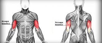
The forearms consist of many relatively large and small muscles, among which the largest is the brachyradialis (brachioradialis muscle), which practically determines the volume of the forearm. In addition, from the point of view of bodybuilding, the flexors and extensors of the wrist, as fairly large muscle groups, are of interest. The forearm also includes finger flexors and extensors, pronators, supinators and other muscles, the training of which is beyond the scope of this article. The flexors are on the inside of the forearm, and the extensors are on the outside.
Exercises[edit | edit code]
Samson's isometric exercises for forearms
- forearm training using a manual expander
- forearm training using a manual expander
- Overhand grip barbell curl
- Barbell Wrist Curl
- Wrist extension
Overhand grip barbell curl[edit | edit code]
It is performed similarly to the biceps curl, only the barbell is taken with an overhand grip. The main load falls on the brachiradialis; in addition, the brachialis and biceps work. The weight of the barbell in this exercise will be much less than with conventional biceps curls, since the brachyradialis is much weaker than the biceps.
Zottman Curls[edit | edit code]
This exercise uses the same muscles as the previous one, only it is performed with dumbbells. At the bottom, the dumbbells are held, as in hammer lifts, and as they move upward, the hands turn palms down (pronation). Then the dumbbells are lowered down, repeating the entire movement in reverse order.
Barbell wrist curl[edit | edit code]
Sit on a bench, place your forearms between your knees on the same bench (or on your knees) so that your hands hang over the edge. Take the barbell with an underhand grip and lift your hands with the barbell up. Return to the starting position. You need to do 15 - 20 such repetitions. The exercise is designed to develop the wrist flexors. It can also be performed with dumbbells.
Standing wrist curl with a barbell behind your back[edit | edit code]
You need to stand with your back to the barbell rack, palms facing back. Take the barbell from the racks and perform flexion-extension of the hands without bending your elbows. The exercise also develops the wrist flexor muscles.
Wrist extension[edit | edit code]
Sit on a bench, place your forearms on the bench or on your knees so that your hands hang freely from them, palms down. Take the barbell with an overhand grip and lift your hands up, then lower them down. This exercise works the wrist extensors.
Rotation of the handle with a suspended load[edit | edit code]
Many gyms have a simulator for developing the muscles of the forearms - a rotating handle, to which a load of different weights can be attached to a cable. You need to grab this handle with an overhand grip and rotate it, winding the cable around the handle, until the load is completely lifted up. Then, rotating in the opposite direction, lower the load into place. You can rotate the handle in two ways - towards yourself and away from you. In the first case, the exercise involves the forearm extensors, in the second - the flexors. In addition, this exercise is very effective for developing grip strength.
General recommendations for performing these exercises[edit | edit code]
Since the forearms are constantly involved both when performing exercises on other muscle groups and in everyday life, their resistance to stress is quite high. Therefore, they belong to the so-called “difficult” muscles and their development is a rather labor-intensive process.
A set of exercises for developing the muscles of the forearms should be performed 2 times a week, each exercise in three approaches, 15 - 20 repetitions in each approach, until failure. Before training the forearms, they need to be thoroughly warmed up and warmed up in order to prevent injury. It is undesirable to allow full stretching of the muscles at the end points of the amplitude.
As part of a training split, it is best to “pump up” your forearms on the day of arm training and on the day of back training. They need to be trained at the end of the workout, after working the arms and back, respectively. Otherwise, full training of the arms and back will be impossible.
2-3 days should pass between forearm workouts, otherwise they will not have time to recover, which can lead to chronic pain in the wrist area. It is advisable to include at least three of the exercises presented above in forearm training - one for the brachyradialis, one for the flexors and one for the extensors of the wrists. It is advisable to change the choice of exercises and the order of their implementation from workout to workout.